INTRODUCTION
 Lung cancer is a very prevalent cancer type and pathological imaging assessment is an important tool for diagnosis.
Previously, pathological imaging reading is done by human doctors.
However human evaluation is objective and labor-intensive.
In addition, pathological images may contain important prognosis and treatment response information that cannot be reliably understood by humans.
Therefore, it is a very important to create machine learning algorithms to automatically recognize pathological imaging data and correlate the automatically
retrieved features with patient phenotype.
Lung cancer is a very prevalent cancer type and pathological imaging assessment is an important tool for diagnosis.
Previously, pathological imaging reading is done by human doctors.
However human evaluation is objective and labor-intensive.
In addition, pathological images may contain important prognosis and treatment response information that cannot be reliably understood by humans.
Therefore, it is a very important to create machine learning algorithms to automatically recognize pathological imaging data and correlate the automatically
retrieved features with patient phenotype.
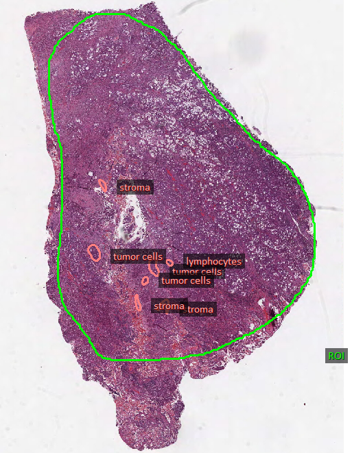
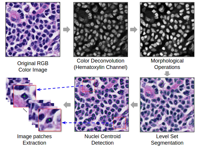
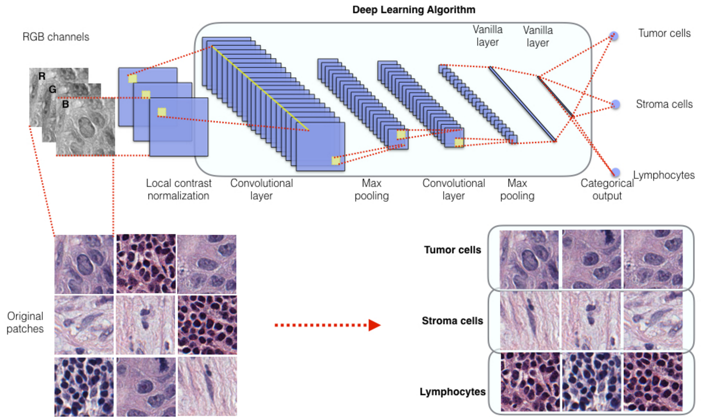
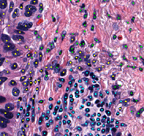
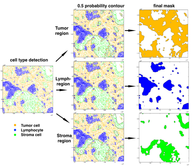
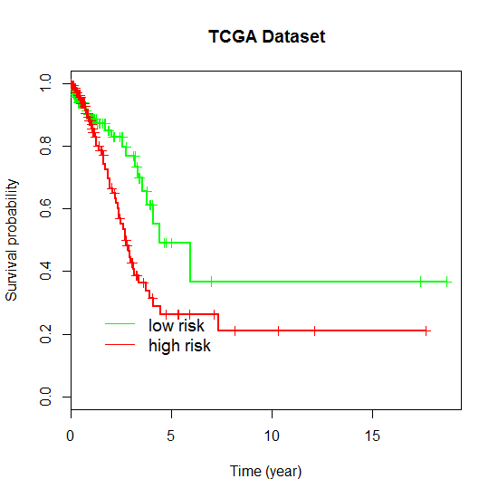
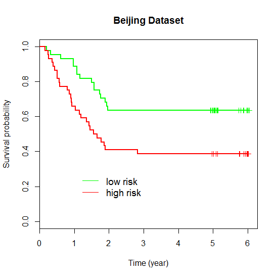
In this study, we obtained pathological imaging slides and prognosis information of patient from TCGA lung adenocarcinoma project (TCGA dataset), the NLST project (NLST dataset), MDACC Lung Spore project (Spore dataset), and the patients from National Cancer centre/Cancer Hospital of Chinese Academy of Medical Sciences, China (Beijing dataset). We developed an imaging analysis pipeline involving image segmentation and convolutional neural network to recognize the cell types in pathological imaging data. And we showed that the extracted cell type-level features by this pipeline are predictive of lung adenocarcinoma prognosis by training a coxph model. This is an early effort to build image analysis pipelines to recognize cell types on lung cancer pathological images and predict patients’ prognosis based on cell type-level features. This approach can potentially be extended to other types of tumor and may be able to be applied to clinical practices in the future.
KEYWORDS: deep learning, lung adenocarcinoma, pathological imaging, prognosis, tumor heterogeneity