Quantitative Biomedical Research Center · UT Southwestern Medical Center
Xenium
Xenium tissue microarray workflow and compact TMA services
The Cai Lab and the UTSW Histopathology Core provide Xenium-compatible tissue microarray (TMA)
construction and spatial transcriptomics services. We build compact TMA recipient
blocks from investigator-provided FFPE donor blocks, sized for the Xenium capture area, and
support the full workflow — from FFPE block contribution and H&E review through ROI selection,
TMA construction, Xenium data processing, annotation, and controlled core-level data return.
The Cai Lab and the UTSW Histopathology Core support complementary parts of a Xenium-compatible
tissue microarray workflow. The Cai Lab coordinates a lung cancer model spatial atlas workflow
around H&E review, FFPE block contribution, ROI selection, TMA planning, Xenium data processing,
annotation, and controlled data return. The Histopathology Core provides compact TMA recipient
blocks and full tissue microarray construction designed for Xenium capture-area utilization.
500+FFPE donor blocks aggregated
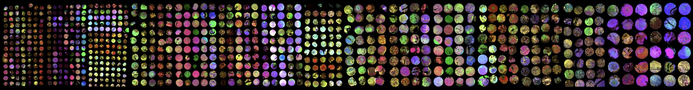
784cores profiled in the current atlas
16TMAs arranged on Xenium slides
21M+cells generated for analysis
Cai Lab workflowLung cancer Xenium TMA atlasFor collaborators contributing H&E images, FFPE blocks, model metadata, or spatial data questions.
The NCI-funded initiative connects community-contributed lung cancer model materials with pathology review,
compact tissue microarrays, and Xenium spatial transcriptomics.
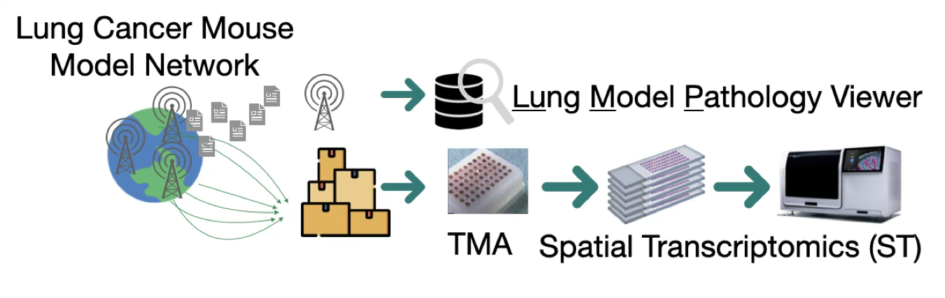
From lung cancer model network to spatial atlas
We are building pathology and spatial transcriptomics resources for lung cancer preclinical model
cross-comparison. The project collects H&E images for import into the Lung Model Pathology
Viewer and FFPE samples for compact TMA construction and subsequent Xenium spatial transcriptomics,
following a traceable image-to-TMA-to-data workflow.
The workflow is especially relevant for investigators working with lung cancer patient tumors and
diverse in vivo preclinical models of lung cancer, including genetically engineered mouse models,
carcinogen-induced tumors, spontaneous tumors, syngeneic and orthotopic models, xenografts, PDXs,
and humanized mouse models.
Workflow description
This workflow connects pathology images, region-of-interest selection, TMA extraction
plans, Xenium profiling outputs, computational annotation, pathologist-guided review, and
contributor-specific data return.
Digital H&E image organization through iViewer, developed by Xiao lab.
ROI registration and TMA planning linked from donor block to TMA core to Xenium output.
Cross-species spatial profiling strategy for human tumors, mouse tumors, and human tumor-in-mouse models.
Core-level data packages suitable for controlled collaborator review and later atlas integration.
How the workflow runs
1. IntakeShare study goals, FFPE block inventory, H&E images, and model metadata.
2. ReviewRegister H&E images and select pathology-informed regions of interest.
3. PlanCreate a compact TMA layout matched to tissue type, core diameter, and Xenium capture area.
4. ProfileConnect TMA construction, Xenium runs, and core-level data extraction.
5. ReturnPackage images, annotations, QC, and controlled interactive review for collaborators.
Workflow and atlas collaboration:Ling.Cai@UTSouthwestern.edu
Use this contact for study design, data processing, annotation, controlled data return, and lung cancer model atlas collaboration.
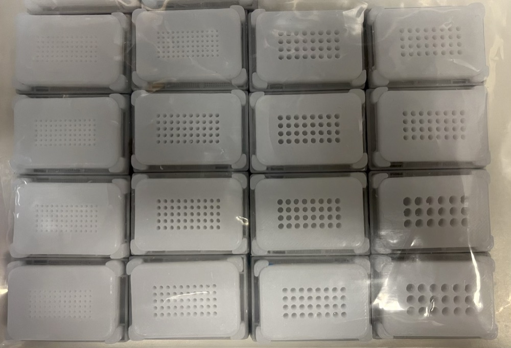
UTSW Histopathology Core serviceCompact Xenium-compatible TMAsFor investigators needing TMA recipient blocks or full tissue microarray construction.
Mold-casted compact TMA recipient blocks
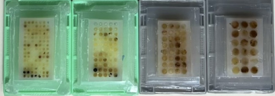
TMA sections placed within Xenium capture areas
Autoarrayer-constructed TMA after sectioning for Xenium
Compact TMA layouts
New Xenium-focused layouts are in development, including 0.6 mm core options
and denser arrangements for larger core sizes. Final layouts will be posted after
mold testing.
1 mm7 x 15 = 105
1.5 mm5 x 11 = 55
2 mm4 x 8 = 32
3 mm3 x 6 = 18
Xenium-Compatible TMA Construction Service
Service options
The UTSW Histopathology Core can support compact TMA recipient blocks and full tissue
microarray construction from investigator-provided FFPE donor blocks.
Compact recipient blocks designed around Xenium capture-area planning.
Full TMA construction for 1 mm, 1.5 mm, 2 mm, and 3 mm tissue cores.
TMA layouts intended for downstream Xenium spatial transcriptomics, morphology imaging, and core-level analysis.
Use this contact for recipient blocks, compact TMA construction, and Histopathology Core service questions.
Please route service requests for TMA recipient blocks or full TMA construction to the
UTSW Histopathology Core.
Please route Cai Lab collaboration requests to Ling Cai for study design, atlas
participation, spatial data processing, annotation, and controlled data-return planning.