Project 1

Summary


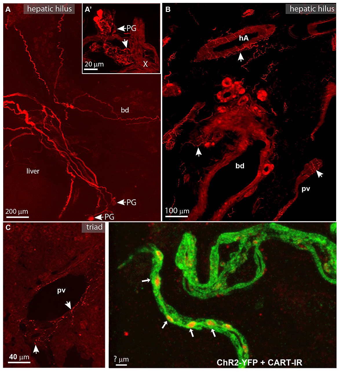
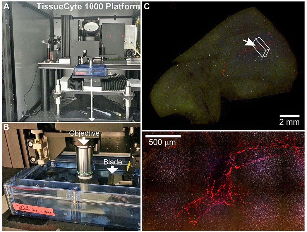
The goal of Project 1 is to generate three-dimensional and neurochemical maps of vagal innervation of the mouse liver. We will also systematically compare the anatomical and neurochemical findings between mice and humans.


Questions


  1. What is the exact topography of the hepatic branch proper and its offshoots?

  2. What are the target cells of the vagal efferents?

  3. What can be sensed by vagal afferents terminating in the liver, gallbladder and around the hilar vasculature? What is the neurochemical profile of the vagal afferents innervating the liver?


Methods


Fluorescently labeled vagus nerve bundles and axons in the liver.
Overview of the automated two-photon platform.
Overview of the automated two-photon platform. A. TissueCyte 1000 consists of a two-photon microscope with an integrated vibratome. B. Agarose-embedded samples are placed in the vibratome and images are collected at the surface of each section. The platform is fully robotized. C and D. Fluorescently labelled axons in the mouse liver.


Team members


Title Name Link
Project Director Laurent Gautron View
Project co-Director Joel Elmquist View
Collaborator Whole Brain Microscopy Facility (Julian Meeks and Denise Ramirez) View
Collaborator Dr. Ronald Bleys, Department of Anatomy, University of Utrecht View